Overview of Our Research
The Wang Lab in the Department of Radiology at UC Davis Health focuses on advancing molecular imaging through the methodological development and clinical translation of parametric positron emission tomography (PET) methods (see a technical overview in Wang et al., 2020). Unlike standard PET, of which the measured signal is commonly a mix of multiple radiotracer states and mainly provides semiquantitative measures, parametric PET offers compartmental resolution—enabling the separate visualization and quantification of individual compartments and their associated tracer kinetics (see YouTube video for a demonstration). As a result, this parametric approach allows for a quantitative, multiparametric characterization of underlying molecular processes. We develop innovative dynamic PET imaging algorithms and advanced tracer kinetic models to drive parametric PET towards higher compartmental resolution, exploring the molecular imaging of biological processes that have been beyond the reach of conventional PET methods.
Current Research Topics
1. Molecular Transport Imaging of Radiotracers![]()
Most radiotracers for PET imaging are utilized to assess their end-product properties (e.g. using [F18]FDG to assess glucose metabolism). The intermediate molecular transport processes of radiotracers across a vascular barrier (e.g., blood-brain barrier) are much less explored but may provide rich information on health and disease. Our lab is developing enabling parametric PET methods to image the molecular transport properties of radiotracers using dynamic imaging and tracer kinetic modeling. We have also successfully applied molecular transport imaging methods to address critical clinical needs in major diseases, such as imaging chronic liver inflammation. [see more details …]
2. Total-Body PET Kinetic Modeling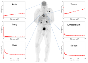
PET parametric imaging has traditionally been limited to single organs, such as the brain or heart, due to the restricted axial field of view of conventional PET scanners, which typically range from 15 to 30 cm. However, the advent of total-body and other long axial field-of-view PET scanners (e.g., the 2-m long EXPLORER) provides ultra-high sensitivity and the ability to simultaneously scan the entire body for dynamic imaging, making total-body multiparametric PET imaging feasible.
Our lab is dedicated to tackling the technical challenges in the emerging field of total-body PET kinetic modeling research using a systemic perspective. These developments may create new methods to help us explore the interactions between different organs and systems in relation to various diseases. For instance, we investigate how the liver, bone marrow, and brain change and interact in cancer patients and in those with metabolic dysfunction-associated steatotic liver disease. [see more details …]
3. PET-enabled Dual-Energy CT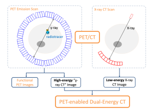
Understanding tissue compositions can be crucial for advancing molecular imaging. This may be done by using X-ray spectral CT imaging, but with critical technical limitations when combined with PET/CT. We have developed a novel concept based on PET/CT physics and advanced image reconstruction algorithms to enable dual-energy CT imaging using combined PET and CT scanning. This PET-enabled dual-energy CT method can add a new dimension of tissue composition information alongside existing PET/CT functional imaging, without the need for costly hardware upgrades or increased radiation exposure. We are currently exploring innovative applications of this technique, particularly in parametric PET imaging of bone marrow. [see more details …]
Funding Acknowledgements:
![]()
![]()


![]()
![]()
and start-up funds from the UC Davis School of Medicine and Cancer Center.