Overview of Our Research
The Wang Lab in the Department of Radiology at the UC Davis Medical Center is focused on the theory and applications of parametric imaging with positron emission tomography (PET) (Wang et al 2020). Our research approach commonly integrates (1) multidimensional data acquisition (e.g., 4D space-time data) with (2) design of computational imaging algorithms (including tracer kinetic modeling, image reconstruction, and machine learning), and (3) discovery of quantitative parametric imaging biomarkers to enable clinically efficient and effective imaging methods in human diseases. By developing advanced PET parametric imaging algorithms and translating them for clinical applications, we can make molecular imaging better, cheaper, safer, and more informative in the clinic.
Current Research Topics
1. Molecular Transport Imaging of Radiotracers![]()
Most radiotracers for PET imaging are utilized to assess their end-product properties (e.g. for assessing glucose metabolism with F18-FDG). The intermediate molecular transport processes of a radiotracer (e.g., the rate of transport across cell membranes) are much less explored but they may provide rich information on health and disease. A major focus of our lab is to develop parametric PET methods to enable the measurement of the molecular transport properties of a radiotracer for single-tracer multiparametric imaging. The enabling techniques include (a) dynamic PET imaging with a high temporal resolution, e.g., using high-performance scanners and/or advanced image reconstruction methods (e.g., the kernel methods Wang & Qi 2015, Wang 2019, Li & Wang 2022, Li et al 2023) and (b) tracer kinetic modeling that allows more accurate estimation of the transport rates of a radiotracer in different organs, for example, for the liver (Wang et al 2018, Zuo et al. 2019 ), the lungs (Wang et al. 2023, Wang et al 2024), the heart (Zuo et al 2020), and the brain (Zhu et al 2024). We have translated our parametric PET methods of molecular transport imaging to address unmet clinical needs in major diseases, such as for assessing liver inflammation in metabolic dysfunction-associated steatohepatitis (Wang et al 2018, Sarkar et al 2019, Sarkar et al 2021), measuring myocardial blood flow in heart disease (Zuo et al 2021), and evaluating neurocognitive changes in systemic diseases.
2. Total-body PET Kinetic Modeling
Parametric imaging using dynamic PET and tracer kinetic modeling has traditionally been limited to single organs like the brain or heart due to the restricted axial field of view of conventional PET scanners (15-30 cm). However, whole-body PET kinetic quantification is essential to study metastatic cancer, organ crosstalk in systemic diseases, or systemic response to therapy. Our team at UC Davis has significantly contributed to whole-body PET parametric imaging through the development of direct reconstruction algorithms using optimization transfer and new kinetic modeling methods that are simplified for clinical use. Our Nested EM algorithm (Wang & Qi 2010) and Relative Patlak Plot (Zuo et al 2018) have been integrated into commercial PET scanners for whole-body parametric imaging by major manufacturers. Nonetheless, whole-body parametric imaging with conventional scanners still has a relatively low temporal resolution while covering the whole-body axial range, mainly limiting to macro-parametric imaging.
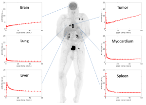 The advent of total-body PET scanners such as the EXPLORER offers ultrahigh sensitivity and simultaneous coverage of the entire body for dynamic imaging. Our lab is at the forefront of exploring the potential of total-body dynamic PET for both macro-parametric imaging and micro-parametric imaging (Wang et al 2021). We are developing methods to address technical challenges, such as optimizing the use of image-derived input function through voxelwise time-delay correction and model selection (Wang et al 2022) and pushing a higher compartmental resolution using high-frame-rate imaging (Wang et al 2024). We have also conducted clinical translation studies that demonstrate the advantages of total-body multi-parametric PET in studying COVID-19 (Wang et al 2023), cancer (Wang et al 2022, Wang et al 2024), and fatty liver disease.
Educational lecture slides on Total-body PET Kinetic Modeling at Total-Body PET 2021-2022, Edinburgh, Scotland.
3. PET-enabled Spectral CT
We explore a novel concept based on PET/CT physics and advanced image reconstruction algorithms to enable dual-energy or multi-energy spectral CT imaging using combined PET and CT scanning. This PET-enabled spectral CT method can add a new dimension of tissue composition information on top of existing PET/CT functional imaging without costly hardware upgrades or increasing radiation exposure. Novel applications of this technique are being explored for PET parametric imaging.
Funding Acknowledgements:
![]()
![]()


![]()
![]()
and start-up funds from the UC Davis School of Medicine and Cancer Center.